
Laboratory Tests and Ancillaries
 Polycystic Ovarian Syndrome_Diagnostic 1
Women with polycystic ovarian syndrome present with a normal to mildly elevated prolactin level, a normal to moderately elevated testosterone level, a normal to mildly reduced FSH level, and a generally moderately elevated LH level. The LH/FSH ratio is elevated at >2.5. A urine pregnancy test should be obtained from every patient in the reproductive age group reporting for menstrual abnormalities. Midluteal serum progesterone may be measured to document potential anovulation or ovulatory dysfunction in polycystic ovarian syndrome patients with regular menses. Biochemical hyperandrogenism may be evaluated by measuring total or free testosterone or free androgen index using chromatography assays. If total or free testosterone levels are not increased, consider measuring androstenedione, DHEAS, and SHBG. If results are elevated, consider an androgen-secreting tumor; normal androgen levels do not rule out polycystic ovarian syndrome.
Serum anti-mullerian hormone (AMH) may be used for defining polycystic ovarian morphology (PCOM) in adults as an alternative to pelvic ultrasound but should not be used as a single test to diagnose polycystic ovarian syndrome. Serum AMH is significantly elevated in women with polycystic ovarian syndrome compared with normal ovulatory women. Determination of AMH is not necessary for polycystic ovarian syndrome diagnosis in patients with irregular menstrual cycles and hyperandrogenism.
Patients should undergo the following lab tests to exclude the corresponding conditions: Glucose tolerance test for DM; fasting lipid panel for dyslipidemia; alanine aminotransferase and aspartate aminotransferase for hepatic steatosis; corticotropin-stimulated serum 17-α-hydroxyprogesterone for congenital adrenal hyperplasia; total and/or bioavailable testosterone and DHEAS for androgen-secreting tumors of the ovary or adrenal gland; 24-hour urinary free cortisol and creatinine, midnight salivary cortisol, or 1-mg overnight Dexamethasone suppression test for Cushing syndrome; thyroid function studies (eg serum thyroid-stimulating hormone [TSH]) for thyroid disease; serum prolactin for hyperprolactinemia; and FSH level for primary ovarian insufficiency.
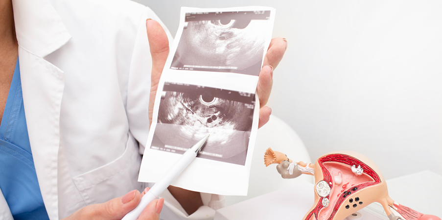
Imaging
Ultrasonography
 Polycystic Ovarian Syndrome_Diagnostic 2
Ultrasonography is used in the diagnosis of polycystic ovarian syndrome in patients who are >8 years past their menarche because a high incidence of multifollicular ovaries is seen in females with a gynecological age of <8 years. Transvaginal ultrasound is the preferred approach if acceptable to the patient and if sexually active. Follicle number per ovary (FNPO) is considered the most effective ultrasound marker to detect PCOM in adults. FNPO, follicle number per cross-section (FNPS) and ovarian volume (OV) are considered accurate ultrasound markers for polycystic ovarian morphology (PCOM) in adults. PCOM criteria should be based on follicle excess (FNPO, FNPS) and/or OV.
Determination of polycystic ovaries in one or both ovaries should have an endovaginal ultrasound findings of ≥20 follicles measuring 2-9 mm in diameter per ovary and/or increased OV (≥10 cm3) on either ovary or ≥10 FNPS in at least one ovary. Transabdominal ultrasound should primarily report OV with a threshold of ≥10 mL or FPNS ≥10 in either ovary due to the difficulty of reliable follicle counting using this approach. If there is a follicle >10 mm in diameter, the scan should be repeated at a time of ovarian quiescence in order to calculate volume and area. The presence of one polycystic ovary is sufficient to provide the diagnosis.
Pelvic
ultrasound is not necessary to diagnose polycystic ovarian syndrome in patients
with irregular cycles and hyperandrogenism, though the complete polycystic ovarian syndrome phenotype
will be identified. In the identification of endometrial abnormalities, routine
screening with ultrasound for endometrial thickness to rule out endometrial
cancer in polycystic ovarian
syndrome patients is not recommended.